I. What is Dental X-Ray
Dental X-ray machines, as an essential imaging tool for modern dentists, are increasingly used in various oral medicine activities such as diagnosis and implantation. The main types of equipment currently in use can be categorized into three types: traditional dental radiography units, panoramic machines, and dental CT scanners. So, what are the differences and advantages of dental CT scanners compared to traditional dental radiography units and panoramic machines?
Dental Portable X-Ray Unit
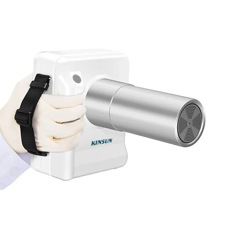
Dental portable x ray units, specifically designed for examining oral teeth, are relatively simple X-ray imaging instruments. They usually capture two-dimensional images of 1 to 4 teeth at a time. However, these images often suffer from significant structural overlap and cover a limited area.
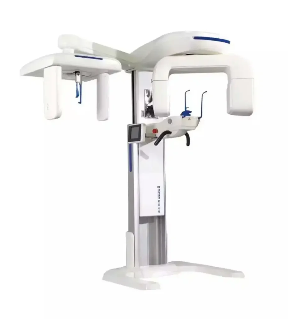
Dental Panoramic machines
Panoramic machines, designed based on the principle of X-ray tomography, are specialized oral X-ray machines capable of capturing a curved plane expansion diagram of the maxillofacial region and the entire dentition in a single shot. On these expansion diagrams, not only can the entire set of teeth be seen, but also parts of the mandible, maxilla, and maxillary sinus, as well as the temporal lobe, temporomandibular joint, and occlusal relationships. These machines play an important role in oral examinations and treatment. However, as they provide two-dimensional images, they are unable to depict three-dimensional structures. Obstructions and shadow effects between different structures are difficult to avoid completely. Dimensions in the buccolingual direction are not displayed, which may lead to missed root canals or misjudgment of buccolingual curvature of the root canal, potentially causing diagnostic errors.
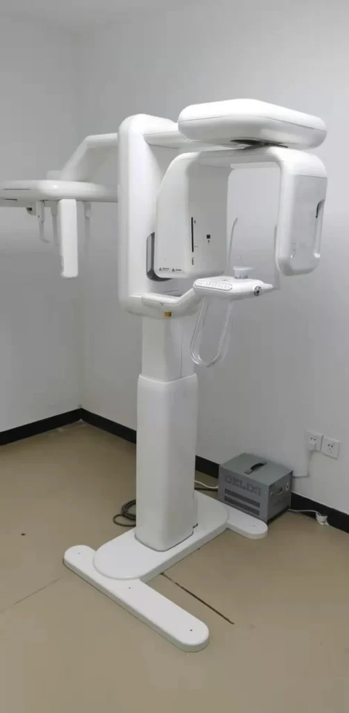
Dental CBCT
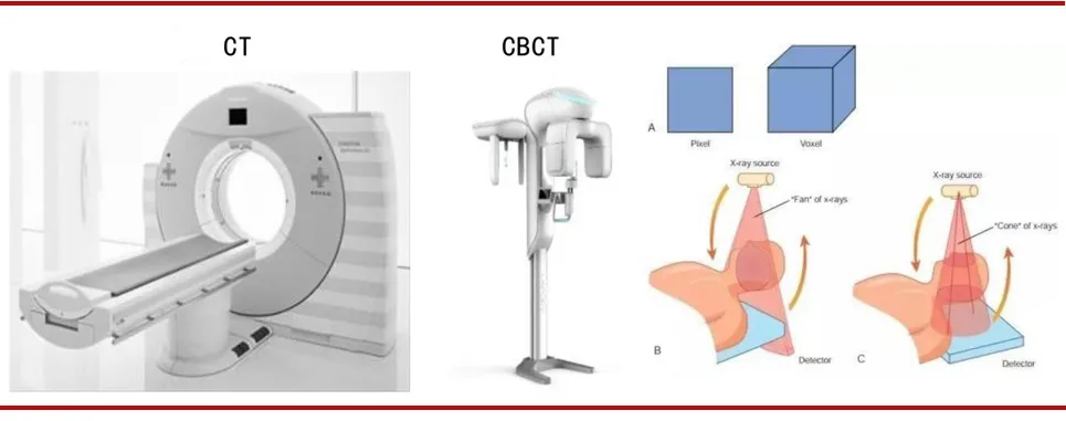
Dental CT, also known as Cone Beam Computed Tomography (CBCT), operates on the principle of using a cone-shaped beam of X-rays. The rays, after passing through the patient, are captured by a flat panel detector. During scanning, the X-ray generator rotates around the subject, collecting data that is then reconstructed in a computer to create three-dimensional images. CBCT allows for a comprehensive 360° three-dimensional observation of the bone density of the dental arch, as well as the height and width of the alveolar bone. It can reconstruct the structure of the entire craniofacial region, measure the structure of the maxillofacial bones, and recreate detailed anatomical details. CBCT is widely used in various fields such as oral and maxillofacial surgery, orthodontics, dental implantology, periodontics, orthognathic surgery, temporomandibular joint disorders, and otorhinolaryngology.
II. Advantages and Disadvantages of CBCT
Advantages and disadvantages of CBCT (Cone Beam Computed Tomography) in oral and maxillofacial imaging diagnosis:
Advantages:
- Three-dimensional observation, including axial, coronal, and sagittal views.
- Images reflect the true size of structures.
- Scanning can be done in standing or sitting positions.
- Isotropic voxels.
- Two-dimensional image observation, including reconstructed curved panoramic planes, and cranial anterior-posterior and lateral views.
- High spatial resolution, suitable for observing trabecular bone structure, root canal structure, periodontal membrane, etc.
- Lower radiation dose.
- Relatively less interference from metal artifacts.
- Relatively simple equipment and easy operation.
- Compatible with DICOM format for storage and transmission, and can be processed with third-party software.
Disadvantages:
- The low-density resolution provides less soft tissue information.
- Limited field of view, varying among different brands and models of machines.
- Increased image noise due to scatter radiation, resulting in a low signal-to-noise ratio.
- Longer projection time, leading to significant motion artifacts.
- Artifacts at the edges of the field of view.
- Inability to use CT values to evaluate tissue density.
As the digitization of the oral industry becomes increasingly prevalent, oral CBCT, as an important part of the digital oral industry, is gradually becoming the mainstream oral imaging equipment. Compared to traditional spiral CT, it has advantages like higher spatial resolution, shorter data acquisition time, lower exposure dose, and lower shooting cost.
The advantages of oral CBCT (Cone Beam Computed Tomography) are significant.
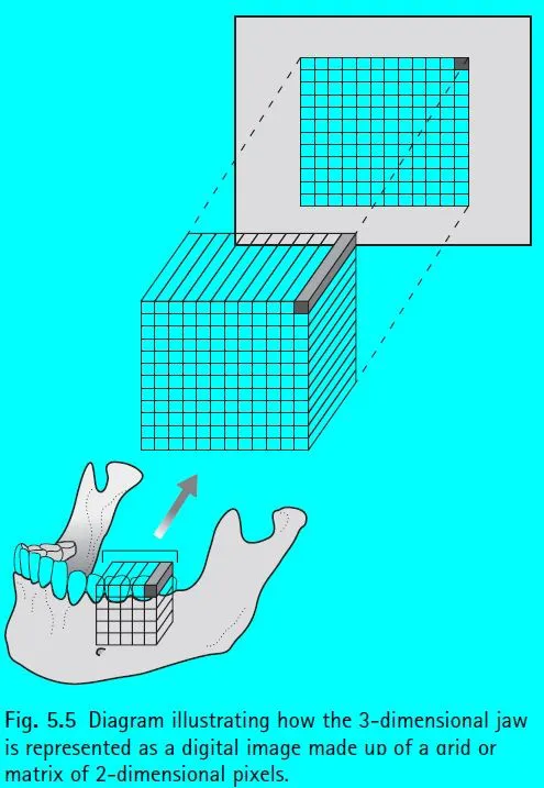
Oral CBCT, short for Cone Beam Computed Tomography, is a type of imaging equipment that reconstructs computed tomography images using a cone-shaped X-ray beam. Its principle involves the X-ray generator emitting a lower dose of radiation (usually around 10 milliamperes tube current) while rotating around the subject in a circular digital projection. The data acquired from multiple digital projections (ranging from 180 to 360, depending on the product) around the subject are then “intersected” and “reconstructed” in a computer to produce three-dimensional images.
The projection principle for data acquisition in CBCT is entirely different from that of traditional spiral scanning CT. Compared to traditional oral imaging methods, CBCT can provide precise three-dimensional structural information. The projection data in traditional spiral scanning CT is one-dimensional, and the reconstructed image data is two-dimensional. The three-dimensional images are composed of multiple two-dimensional slices stacked together, which often results in significant metal artifacts in the images.
CBCT’s use of a cone-shaped X-ray beam significantly improves the efficiency of X-ray utilization. A full 360-degree rotation is sufficient to acquire all the original data needed for reconstruction. Moreover, using a flat panel detector to collect projection data speeds up the data acquisition process.
Due to differences in imaging principles, compared to traditional spiral CT, CBCT (Cone Beam Computed Tomography) boasts several advantages such as higher spatial resolution, shorter data acquisition time, lower exposure dose, and reduced shooting costs.
The application of CBCT is also very broad, encompassing various sub-disciplines within dentistry. These include areas such as implantology, orthodontics, oral and maxillofacial surgery, endodontics, periodontics, and temporomandibular joint disorders.
III. Best Dental Cone Beam Brand
The main brands of CBCT on the market include Germany’s Kavo, Germany’s Sirona, Italy’s New Tom, South Korea’s Vatech, etc.
World CBCT Brand
- Dentsply Sirona
Founded in 1899, Dentsply is headquartered in York County, Pennsylvania. In 2016, Dentsply and Sirona joined forces to create the new Dentsply Sirona, a company dedicated to building a global digital dental and integrated solutions company. Its business scope includes dental disease prevention, dental restoration, orthodontics, root canal treatment, dental implants, oral restoration, healthcare, etc. According to the latest financial report, the company’s profit for the first fiscal quarter of 2018 was $81.2 million, a 35.79% increase year-over-year; revenue was $956 million, a 6.17% increase year-over-year.

Over 100,000 Dentsply Sirona extraoral imaging devices have been installed in dental clinics around the world. High quality, German standards, product reliability, and ease of operation provide clinic owners with a dynamic tool for integration. Reliable after-sales support and specialized product training further ensure customer satisfaction.
- KaVo Sybron
KaVo Group is a designer and manufacturer of dental equipment and consumables, headquartered in the United States. Its business scope includes dental medical device products, disinfectants, sales services, and related support businesses. Offering one-stop solutions and products for oral prevention, diagnosis, and treatment, KaVo Group has established research and development centers in Shanghai and manufacturing plants in Suzhou, Jiangsu, and Ziyang, Sichuan, engaged in researching and manufacturing high-quality products, committed to providing customers with efficient and safe products and services. KaVo Group offers comprehensive large-scale products and services in oral imaging diagnosis, oral restoration, orthodontics, and dental implants, providing professional solutions and services to dentists and patients.
Its CBCT imaging series features technologies such as ORTHOselect™ maxillofacial self-locking shooting technology, ORTHOfocus™ maxillofacial focusing technology, ORTHOceph™ maxillofacial independent projection technology, Low Dose Technology™ low-dose micro-light technology, etc.
- NewTom from Italy
QR (Qualitative Radiology) s.r.l, based in the famous city of Verona, Italy, is the inventor of the world’s first dental cone-beam CT and a market pioneer in this field, with over a decade of experience in manufacturing and R&D of cone-beam CT. Its main products are the third-generation horizontal cone-beam CT – NewTom 3G, and the vertical cone-beam CT – NewTom VG.

Cone-beam computed tomography (CBCT) is widely used in oral and maxillofacial surgery, orthognathic surgery, orthodontics, implantology, periodontics, conservative dentistry, pediatric preventive dentistry, and otolaryngology for imaging diagnosis and analysis.
In April 2007, QR s.r.l (NewTom Dental) became a subsidiary of the American AFP Imaging Corporation (OTCBB: AFPC), a company listed on the American OTC stock market. DENT-X and EVA are the main brands of its dental product series. In addition to dental products, AFP also produces other medical and pet imaging products. AFP and its subsidiaries are involved in technical research, product development, and the production and distribution of their medical, dental, and veterinary products.
China’s Leading CBCT Company
1. BonDent

Established in Hong Kong in 1986 and moved its production line to Shenzhen in 1992, Modern Dental is primarily engaged in the production and distribution of denture materials. It is a leading global manufacturer of dentures, divided into two major categories: fixed dentures and removable dentures. Fixed denture materials include crowns and bridges; removable denture materials include removable dentures. In 2017, the company’s revenue was approximately 2.181 billion Hong Kong dollars, an increase of 32.8% year-over-year; gross profit was about 1.061 billion Hong Kong dollars, up 20.5% year-over-year.
2. Beijing Largev
Originating from Tsinghua University, Beijing Leadman Biochemistry is a high-tech company providing advanced medical imaging products and services. It possesses industry-leading core technologies in high-precision CBCT imaging, artifact correction, and dose control. Currently, it has several multifunctional oral CBCT products such as Smart3D and HiRes3D, both of which have obtained CFDA registration certification.
3. Meyer
Founded in 2000, Meya Optoelectronics is a high-tech enterprise specializing in optoelectronic detection and grading equipment and its application software development. Its main business includes color sorters, oral CT diagnostic machines, and industrial inspection equipment.
The company started obtaining approvals for oral CT (dental X-ray CT diagnostic machines) in 2012 and began marketing its equipment. In 2013, only 61 oral CT units were sold, generating revenue of 22.13 million yuan; in 2014, about 150 oral CT units were sold, with revenue of 48.35 million yuan; in 2015, around 250 units were sold, earning 75.11 million yuan in revenue; in 2016, nearly 600 oral CT units were sold, achieving sales revenue of 165 million yuan, a year-over-year increase of 120.22%; in 2017, the company sold about 1000 oral CT units, with revenue of 260 million yuan, a 57.28% increase from the previous year, and its domestic market share was expected to be over 30%.
IV. Fundamentals of Radiation Protection for Dental X-Ray Machines
Consideration of X-Ray Generation from a Clinical Perspective
X-rays and gamma rays have a wavelength range of 10 nm to 0.01 pm, with a photon energy range of 124 eV to 124 MeV. From a clinical perspective, the generation of X-rays involves the following steps:
- Electrically heating a filament to produce an electron cloud around it.
- The high voltage between the anode and cathode of the X-ray tube accelerates the electrons around the anode to a very high speed.
- Focusing devices concentrate the electron beam onto a target point.
- Electrons bombard the target point and suddenly stop moving.
- Most of the energy lost by the electrons stopping (99%) is converted into heat, while a small part (1%) is converted into X-rays.
- When heating stops, the heat is absorbed and blocked by the surrounding copper and oil.
- X-rays are emitted from the target point in all directions, with a small portion of the rays being directed in a specific direction through guiding equipment to form the X-rays used in radiographic examinations.
About Radiation Measurement
The units of radiation dose measurement include Radiation Absorbed Dose (D), Equivalent Dose (HT), Effective Dose (E), Collective Effective Dose or Collective Dose, Dose Limits, and Dose Rate.
1. Radiation Absorbed Dose (D): This refers to the energy absorbed per unit mass of tissue from radiation, which can be measured using a dosimeter.
- Standard unit: Joules per kilogram (J/kg)
- Special name: Gray (Gy)
2. Equivalent Dose (HT): This allows for comparison of the radiobiological effectiveness (RBE) of different types of radiation. It uses a radiation weighting factor (WR) to represent the biological effect of different types of radiation on different tissues. The unit for the equivalent dose (HT) provides a comparison of the effects of different types of radiation in a specific tissue. For example:
- X-rays, gamma rays, beta rays: WR = 1
- Fast neutrons (10keV-100keV) and protons: WR = 10
- Alpha particles: WR = 20
- The equivalent dose in a specific tissue is calculated as:
HT = Radiation Absorbed Dose (D) × Radiation Weighting Factor (WR) - Standard unit: Joules per kilogram (J/kg)
- Special name: Sievert (Sv)
3. Effective Dose (E): Allows comparison of the amounts of radiation absorbed by different parts of the body, as some parts are more sensitive to radiation than others. The International Commission on Radiological Protection (ICRP) provides a tissue weighting factor (WT) for each organ or tissue, based on its sensitivity to radiation. The higher the sensitivity, the larger the value. The sum of these values represents the total weighting factor for the entire body.
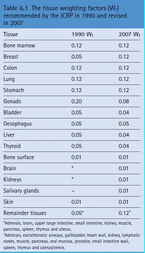
The Effective Dose (E) for the entire body is calculated as the sum of the Equivalent Absorbed Doses (HT) for each tissue multiplied by the Tissue Weighting Factor (WT) of that tissue.
- Special name: Sievert (Sv)
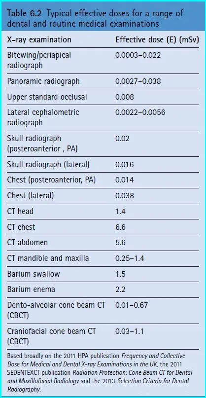
Due to the imprecision of using dose alone, the Effective Dose (E) is commonly used as a descriptor. The Effective Dose can be considered a broad indicator used to assess the risk to health from any kind of radiation exposure to the body. It does not take into account the type of radiation, its energy, or the specific area of exposure. The Effective Doses for common examinations are as follows:
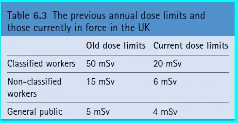
4. Dose Limits
For individuals working with radiation are primarily set with the principle that the health risk to the worker from radiation within these limits should not be greater than the risks from other factors (non-radiation, environmental).
The standards in the United Kingdom are as follows:
According to international standards, taking the annual effective dose of 1 mSv for the general public as an example, one can have 45 periapical radiographs, 26 panoramic radiographs, or 1-2 CBCT scans. If measured by the standards for workers, the above numbers should be multiplied by 20. (All the above calculations are based on the maximum effective dose for each type of examination.)
Tissue Damage Caused by Radiation
Direct damage includes the inability to transmit genetic information; abnormal replication; cell death; and temporary damage (DNA can repair itself). If the DNA of somatic cells is damaged, it may induce tumors. If stem cells are damaged, it could lead to congenital abnormalities.
Indirect damage includes the production of free radicals from the ionization of water by radiation, which indirectly harms cells.
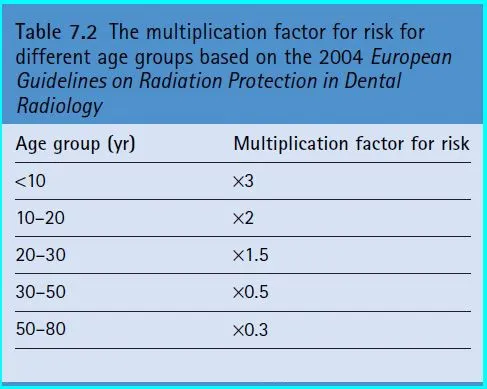
The relationship between risk and age: The younger the individual, the higher the risk; conversely, the older the individual, the lower the risk.
Women’s risk is slightly higher than men’s.
Although the risk of cancer induced by low-dose dental radiographic examinations is very low, the number of people undergoing oral and other radiological examinations is quite large. In the UK, it is estimated that there are 2 million such examinations per year. Among these, there may be about 700 cases of induced cancer, with approximately 10 cases originating from oral radiology examinations.
Hereditary Effects (Genetic Effects):
Primarily based on data from mouse experiments, a dose of 0.5-1.0 Sv can double the probability of natural variations.
Impact on Fetuses:
The dose of radiation used in dentistry is very low, below the threshold for producing tissue effects. However, the occurrence of stochastic effects is not dose-dependent.
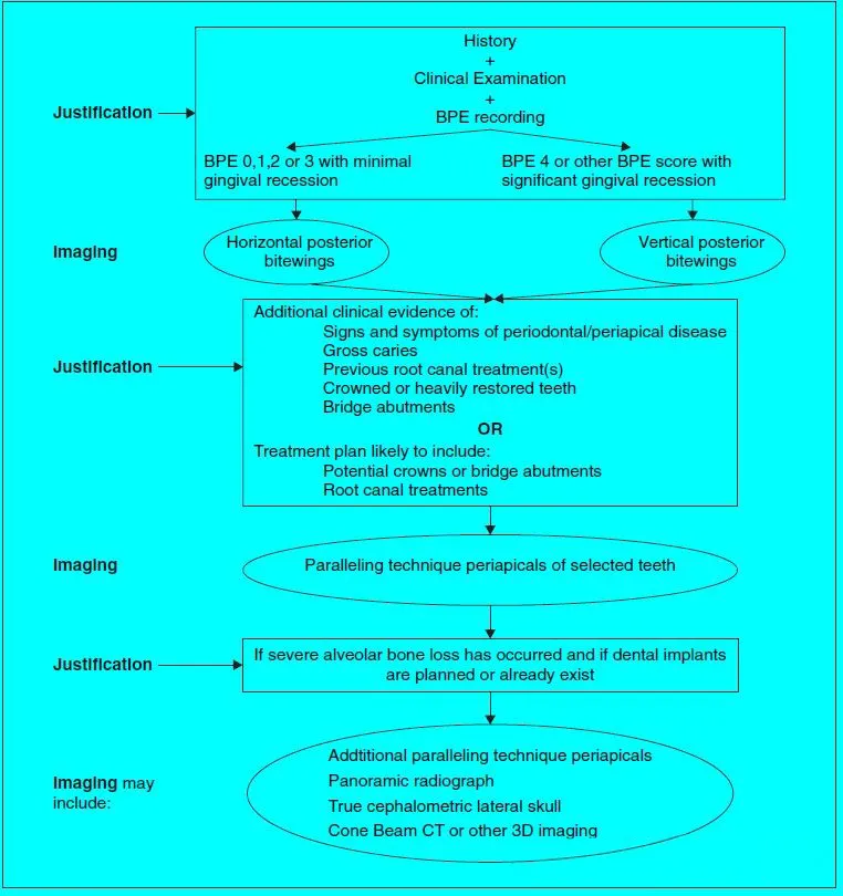
Flowchart of the decision-making process for imaging studies.
Protection for dentists and other staff
Sources of Radiation Exposure:
- Direct exposure, when standing in the path of the radiation beam.
- Reflected from the patient when they are near the patient.
- Leakage of radiation from the X-ray tube.
Basic Principle of Protection: Inverse square law, where the radiation dose decreases in a square proportion with the increase in distance.